
MRI 1.5 BRAIN - ROUTINE
- List Item #1
MRI 1.5 LUMBAR / LUMBO SACRAL SPINE
- List Item #1
MRI 1.5 CERVICAL SPINE
- List Item #1
MRI 1.5 ONE REGION SCREENING
- List Item #1
MRI 1.5 PELVIS
- List Item #1
MRI 1.5 DORSAL SPINE
- List Item #1
MRI 1.5 MRA
- List Item #1
MRI 1.5 MRCP
- List Item #1
MRI 1.5 STROKE PROTOCOL{ MRI+MRA+ASL }
- List Item #1
MRI 1.5 EPILEPSY PROTOCOL+ ASL
- List Item #1
MRI 1.5 MRV BRAIN
- List Item #1
MRI 1.5 BRAIN - ANGIO
- List Item #1
MRI 1.5 HIP
- List Item #1
MRI 1.5 WHOLE ABDOMEN
- List Item #1
MRI 1.5 HEAD
- List Item #1
MRI 1.5 NECK
- List Item #1
MRI 1.5 SELLA/PITUITARY WITHOUT CONTRAST
- List Item #1
MRI 1.5 DORSAL / DORSO LUMBER SPINE
- List Item #1
MRI 1.5 BRAIN - ANGIO AND NECK ANGIO
- List Item #1
MRI 1.5 LEG
- List Item #1
MRI 1.5 WHOLE SPINE
- List Item #1
What is (MRI) Magnetic Resonance Imaging and Why It Matters
Magnetic Resonance Imaging (MRI) is a sophisticated imaging technique used to visualize internal structures of the body in great detail. Unlike X-rays or CT (computed tomography) scans, MRI does not use ionizing radiation. Instead, it employs strong magnetic fields, radio waves, and computer processing to create detailed images of organs, tissues, and other internal structures.
An MRI scanner typically looks like a large tube surrounded by a circular magnet. Patients lie down on a motorized bed that slides into the tube, where the imaging process takes place.
How MRI Works: A Simplified Breakdown
MRI operates on fundamental principles of physics, particularly the behavior of atoms in a magnetic field. Here’s a step-by-step overview of how it works:
Magnetic Alignment:
The human body is mostly made of water, and water contains hydrogen atoms. When a person enters the MRI scanner, the strong magnetic field aligns the hydrogen protons in the body.
Radio Frequency Pulses:
A radio wave pulse is then sent through the body, temporarily knocking the aligned hydrogen protons out of position.
Signal Detection:
When the radio pulse stops, the protons realign themselves with the magnetic field. As they do, they emit signals.
Image Formation:
These signals are detected by receivers in the scanner and processed by a computer to form detailed images of the body’s internal structures.
Different tissues return to alignment at different rates, and this variation is used to distinguish between types of tissues (e.g., muscle, fat, brain tissue) in the resulting images.
Types of MRI Scans
There are several specialized MRI techniques used depending on the clinical need:
T1-Weighted MRI: Provides high-resolution images of anatomy.
T2-Weighted MRI: Highlights fluid-filled areas, useful for identifying swelling or inflammation.
Functional MRI (fMRI): Measures brain activity by detecting changes in blood flow.
Magnetic Resonance Angiography (MRA): Visualizes blood vessels and detects vascular abnormalities.
Diffusion Tensor Imaging (DTI): Maps the diffusion of water in tissues, especially useful in brain imaging.
Applications of MRI
MRI has a broad range of clinical applications, making it a critical diagnostic tool across multiple medical specialties:
1. Neurology
MRI is perhaps most renowned for its role in brain imaging. It helps in diagnosing:
- Brain tumors
- Multiple sclerosis
- Stroke
- Aneurysms
- Brain injuries
- Epilepsy
Neurodegenerative disorders like Alzheimer’s and Parkinson’s disease
Functional MRI (fMRI) is particularly important in brain research, as it shows real-time brain activity, helping scientists and doctors understand brain functions and map critical areas before surgery.
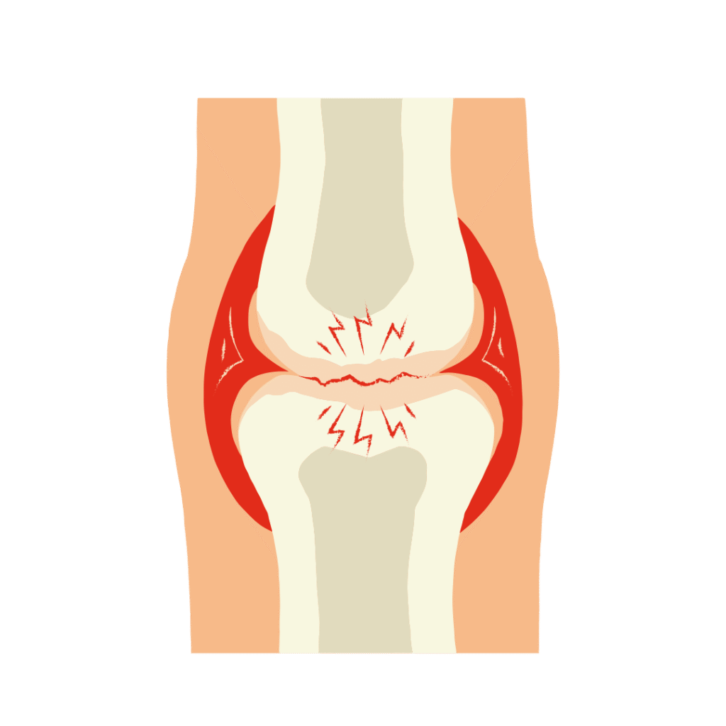
2. Orthopedics
MRI is widely used to evaluate musculoskeletal conditions:
- Torn ligaments and tendons (e.g., ACL tears)
- Cartilage damage
- Joint abnormalities (e.g., arthritis)
- Spinal issues such as herniated discs or spinal stenosis
- Bone tumors
Because it produces high-resolution images of soft tissues, MRI is often more effective than X-rays for detecting certain injuries.
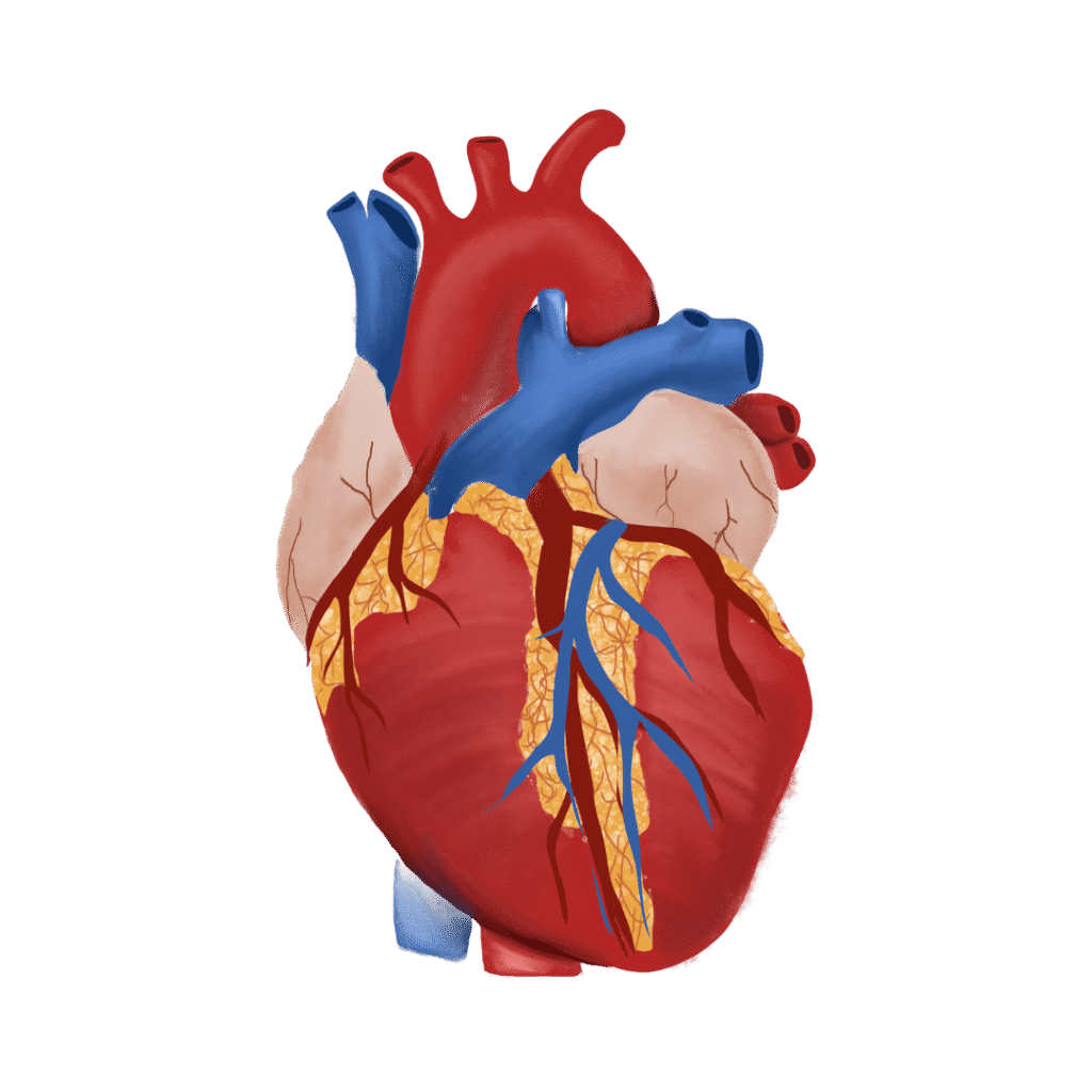
3. Cardiology
MRI of the heart (Cardiac MRI) provides detailed images of the heart’s structure and function. It is used to:
- Evaluate heart size and function
- Detect congenital heart defects
- Assess damage after a heart attack
- Detect pericarditis, cardiomyopathy, or myocarditis
4. Oncology
MRI is crucial in the detection and staging of many types of cancer. It helps determine:
- Tumor size and location
- Whether cancer has spread
- Response to treatment
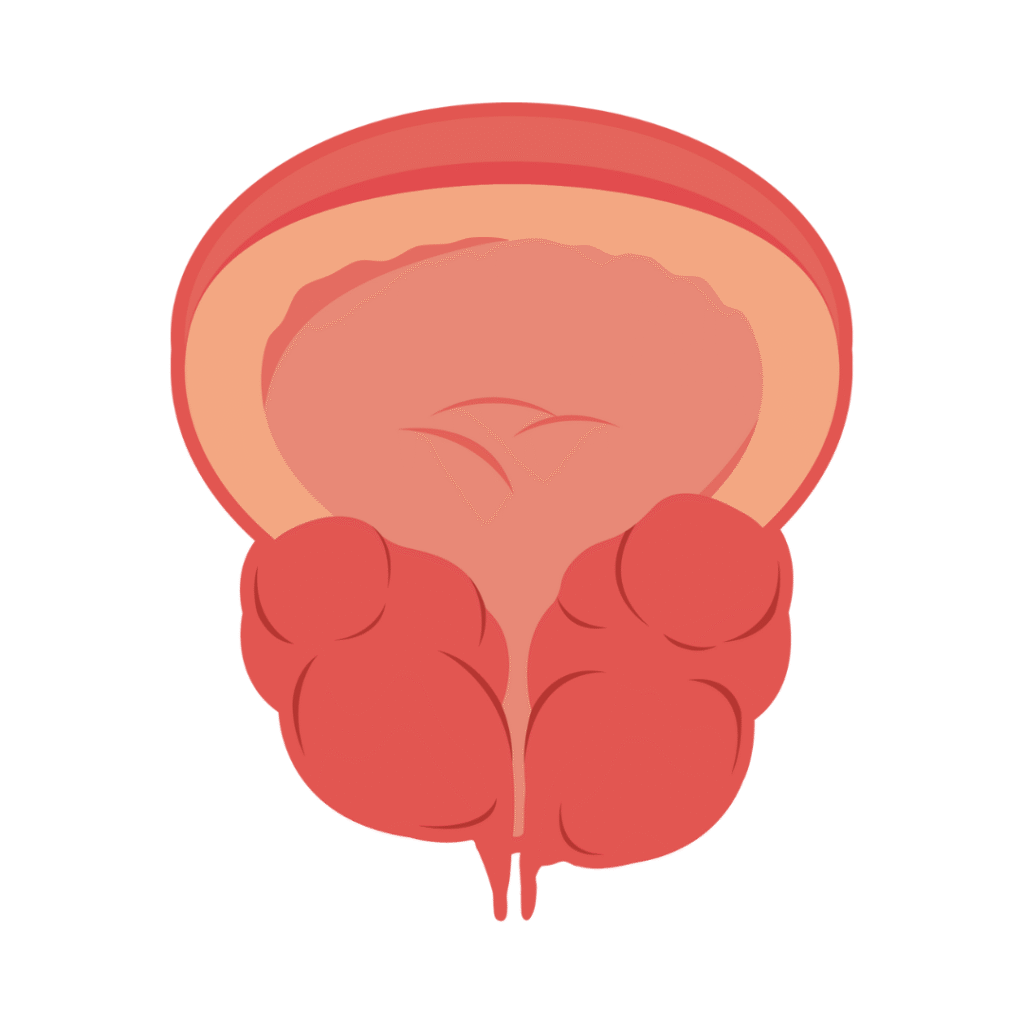
- MRI is often used in diagnosing cancers of the brain, breast, liver, prostate, and spine.
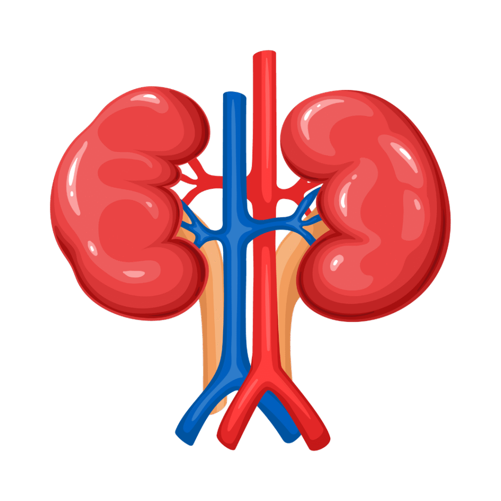
5. Abdominal and Pelvic Imaging
MRI is used to examine internal organs such as the liver, kidneys, pancreas, uterus, and prostate. It is particularly helpful in:
- Detecting liver disease or tumors
- Identifying uterine fibroids and endometriosis
- Diagnosing pelvic pain or infertility
Advantages of MRI
MRI offers several benefits that make it an essential tool in healthcare:
1. Non-Invasive and Painless
MRI does not require surgery or internal probes. Most scans are completely non-invasive and painless, though patients may experience some discomfort from remaining still for an extended period.
2. No Ionizing Radiation
Unlike X-rays and CT scans, MRI does not use ionizing radiation, reducing long-term health risks, especially for children and patients needing frequent imaging.
3. Superior Soft Tissue Contrast
MRI produces higher contrast between different soft tissues than any other imaging modality. This is critical in brain, muscle, and tumor imaging.
4. Detailed and Accurate
MRI allows for early detection of diseases, sometimes before symptoms manifest. Its high sensitivity improves diagnostic accuracy and reduces misdiagnosis.
5. Versatility
MRI can be used to examine nearly every part of the body. It can provide structural, functional, and even metabolic information through various imaging sequences and techniques.
Limitations of MRI
Despite its many benefits, MRI has some limitations:
High Cost: MRI scans are more expensive than X-rays or ultrasound.
Time-Consuming: Scans typically take 30–60 minutes, and some may take longer.
Noise and Claustrophobia: MRI machines can be loud, and enclosed spaces can trigger anxiety in some patients.
Metal Implants: Patients with pacemakers, cochlear implants, or certain metal fragments may not be eligible for MRI.
Movement Sensitivity: Any movement during the scan can distort images, making it less suitable for very young or restless patients unless sedation is used.
Recent Advances in MRI Technology
MRI technology continues to evolve, leading to faster scans, higher image resolution, and greater diagnostic accuracy. Notable innovations include:
3T and 7T MRI Machines: These offer stronger magnetic fields and clearer images.
AI Integration: Artificial intelligence is being used to speed up image reconstruction and enhance interpretation.
Portable MRI: New lightweight MRI systems are being developed for use in remote areas or at the patient’s bedside.
Hybrid Imaging: Techniques like PET-MRI combine functional and anatomical data in a single scan.
The Role of MRI in Medical Research
MRI is not just a clinical tool—it plays a vital role in biomedical research. It helps scientists study:
- Brain connectivity and function
- Effects of new drugs on organs
- Developmental changes in infants and children
- Disease progression and treatment efficacy
fMRI, in particular, has opened new frontiers in cognitive neuroscience, psychology, and behavioral research, allowing us to observe how the brain works in real time.
Why MRI Matters: A Summary
MRI is a cornerstone of modern medicine. Its ability to capture detailed images of soft tissues, organs, and systems without using radiation makes it indispensable for diagnosis, treatment planning, and follow-up.
Key Takeaways:
MRI provides highly detailed, non-invasive images of internal body structures.
It is crucial for diagnosing a wide range of conditions, from brain tumors to joint injuries.
MRI avoids radiation exposure, making it safer for frequent or long-term monitoring.
It plays a vital role in early detection, accurate diagnosis, and personalized treatment.
Advances in technology are making MRI faster, more accessible, and even more powerful.
Conclusion:
In an age where precision and safety are paramount in healthcare, MRI continues to be a technological marvel that saves lives, improves outcomes, and pushes the boundaries of what we know about the human body. Whether it's diagnosing a life-threatening tumor, guiding a complex surgery, or unlocking the secrets of the brain, MRI stands as one of the most important tools in medicine today.
As technology advances and access expands, MRI will undoubtedly remain at the heart of diagnostic imaging for decades to come.


Diabetes
Pregnancy
Thyroid
Liver
Covid
Prostate
Fertility
Bone


Gastro
Cervix
Heart
Kidney
cancer
breast
Vitamins


Tuberculosis (TB)
Anemia
Lungs
Fever
Allergy
Frequently Asked Questions
MRI (Magnetic Resonance Imaging) uses powerful magnets and radio waves to produce detailed images of internal body structures—without using X-rays or radiation.
MRI is commonly used to examine:
Brain and spinal cord
Joints and soft tissues (muscles, ligaments)
Organs like the liver, uterus, prostate, heart
Tumors, injuries, infections, or abnormalities
Yes. MRI is non-invasive and radiation-free, making it safe for most people. However, it is not suitable for patients with certain implants or metal fragments in the body.
Remove all metal objects (jewelry, watches, belts, etc.)
Inform the radiologist if you have pacemakers, metal implants, or pregnancy
You may be asked to fast for a few hours if a contrast-enhanced MRI is planned
Remove all metal objects (jewelry, watches, belts, etc.)
Inform the radiologist if you have pacemakers, metal implants, or pregnancy
You may be asked to fast for a few hours if a contrast-enhanced MRI is planned
Yes, it is generally safe. Minor side effects like nausea or warmth may occur. Please inform the doctor if you have kidney disease or allergies.
An MRI scan typically takes 20 to 60 minutes, depending on the area being scanned. You must lie still during the procedure for accurate imaging.
No pain is involved, but you’ll hear loud knocking sounds from the machine. Earplugs or headphones are usually provided for comfort.
MRI is generally safe during pregnancy (especially after the first trimester), but it’s best avoided unless medically necessary. MRI with contrast is not usually recommended during pregnancy.
Reports are usually available within 24–48 hours. Urgent cases may receive faster reporting if needed.