
X-CHEST PA VIEW
- List Item #1
X-ORTHOPANTOMOGRAM (OPG)
- List Item #1
X-L-SPINE AP & LAT VIEW
- List Item #1
X-C-SPINE AP & LAT VIEW
- List Item #1
X-PNS
- List Item #1
X-KNEE JOINT AP & LAT VIEW (BOTH)
- List Item #1
X-FOOT AP & OBLIQUE VIEW
- List Item #1
MAMMOGRAPHY (DIGITAL)
- List Item #1
X-SHOULDER AP & LAT VIEW
- List Item #1
X-WRIST AP & LAT VIEW
- List Item #1
X-PELVIS AP VIEW
- List Item #1
X-FOOT AP & LAT VIEW
- List Item #1
UROFLOWMETRY
- List Item #1
X-HAND AP & OBLIQUE VIEW
- List Item #1
BONE DENSITOMETRY - SPINE
- List Item #1
X-CHEST AP VIEW
- List Item #1
X-ELBOW AP & LAT VIEW
- List Item #1
X-SHOULDER AP VIEW
- List Item #1
What is an X-ray?
An X-ray is a quick, painless, and widely used medical imaging technique that uses a small dose of ionizing radiation to create pictures of the inside of the body, especially bones and dense structures. X-rays are one of the oldest and most commonly used imaging methods in healthcare.
Why X-ray Matters
X-rays are essential in diagnosing, monitoring, and guiding treatment for a wide range of medical conditions. Their value lies in their speed, accessibility, and effectiveness, particularly in emergencies and basic diagnostics.
Key Uses and Importance
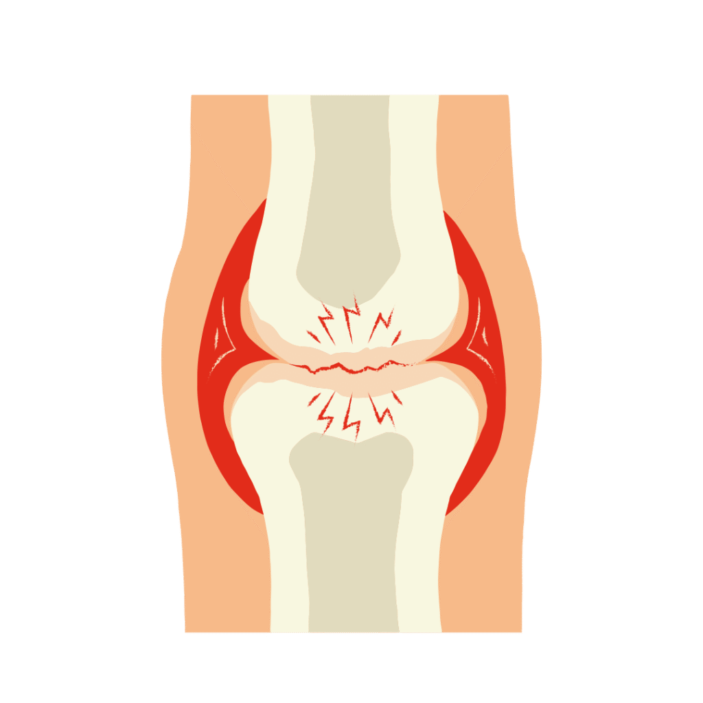
Bone and Joint Evaluation
- Detects fractures, dislocations, and joint abnormalities
- Monitors bone healing after injury or surgery
- Diagnoses arthritis or bone infections
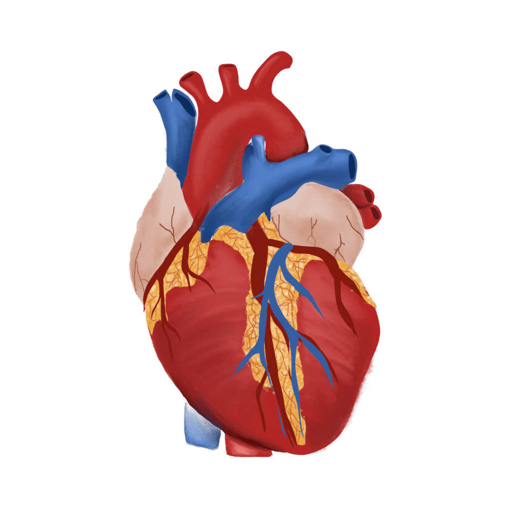
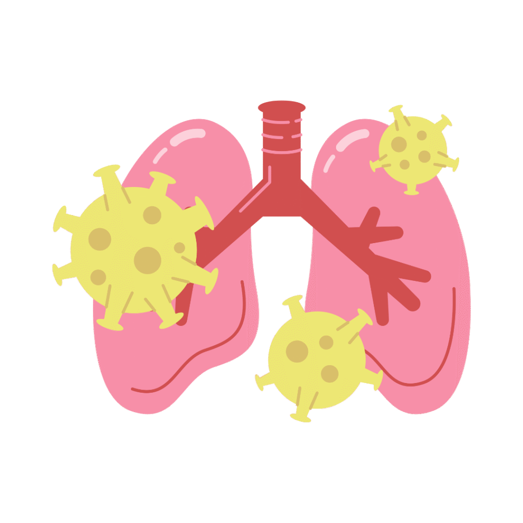
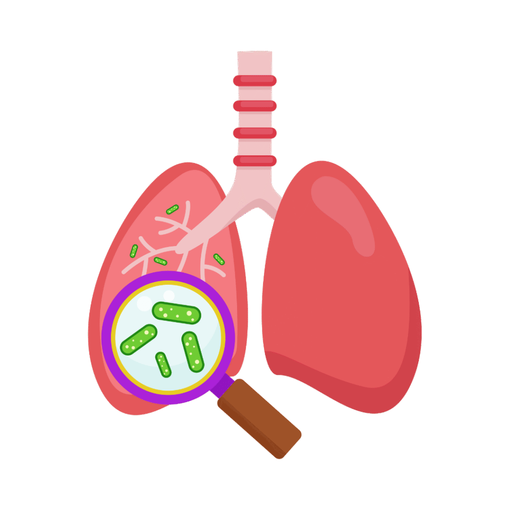
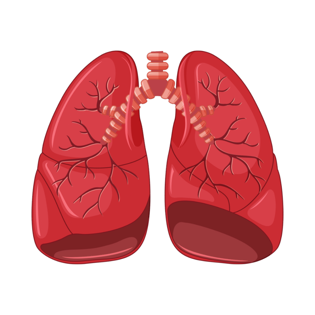
Chest Imaging
- Identifies pneumonia, tuberculosis, lung tumors, or fluid around the lungs
- Assesses heart size and detects conditions like heart failure
Dental Imaging
- Checks for cavities, impacted teeth, jaw alignment, or infections
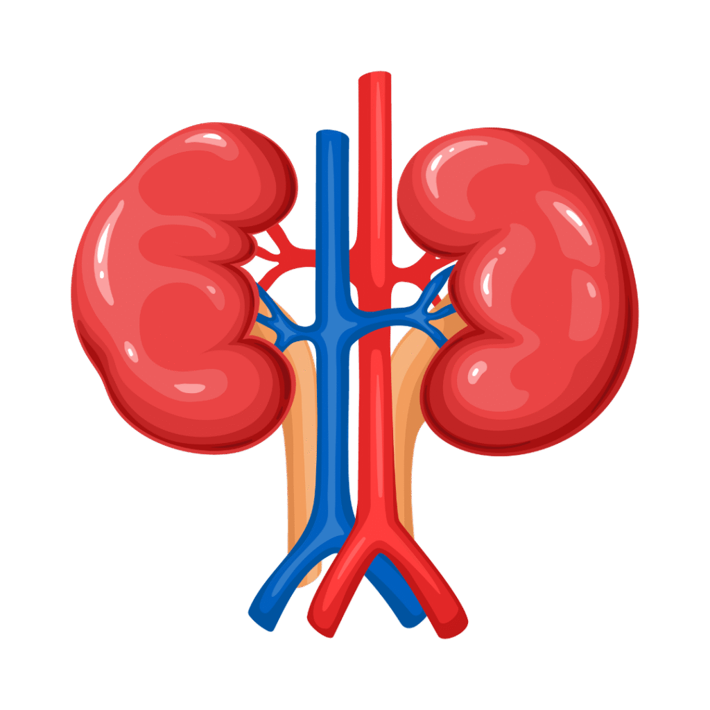
Abdominal X-rays
- Detects intestinal blockages, kidney stones, or swallowed foreign objects
Cancer Detection
- Identifies certain types of tumors or abnormal masses
- Guidance for Medical Procedures
- Used during surgeries or catheter placements (with fluoroscopy, a real-time X-ray)
- Advantages of X-ray
Fast and widely available – Often used in emergency rooms and outpatient clinics - Non-invasive and painless
Effective for bone and chest evaluations
- Cost-effective compared to other imaging methods like CT or MRI
- Limitations and Safety
Exposure to radiation (minimal but cumulative—used cautiously, especially in children and pregnancy) - Less detailed for soft tissues compared to MRI or ultrasound
In summary, X-rays are a fundamental diagnostic tool that provide rapid, critical information—helping doctors make timely decisions, especially in urgent or trauma-related cases.


Diabetes
Pregnancy
Thyroid
Liver
Covid
Prostate
Fertility
Bone


Gastro
Cervix
Heart
Kidney
cancer
breast
Vitamins


Tuberculosis (TB)
Anemia
Lungs
Fever
Allergy
Frequently Asked Questions
An X-ray is a quick, painless imaging test that uses low-dose radiation to produce images of bones, organs, and tissues inside the body.
X-rays are used to:
Detect bone fractures or dislocations
Diagnose chest infections, pneumonia, or lung conditions
Identify joint problems or arthritis
Detect foreign objects or assess digestive tract
Evaluate dental or sinus issues
Yes. The radiation exposure in an X-ray is very low and generally considered safe. However, pregnant women should inform the technician in advance.
In most cases, no special preparation is required. However, for some types (like abdominal X-rays), you may be asked to avoid food or wear a gown.
Wear loose, comfortable clothing and avoid any metal items like zippers, jewelry, or underwire bras that might interfere with the image.
No. X-rays are painless. You may be asked to hold a position briefly, which may be uncomfortable if you're injured, but the procedure itself is quick.
An X-ray typically takes 5 to 10 minutes, depending on the area being examined.
X-rays can scan:
Bones (arms, legs, spine, skull)
Chest and lungs
Abdomen and pelvis
Joints and soft tissues
Teeth and jaw (dental X-ray)
Yes. X-rays are safe for children and commonly used in pediatric care. Special care is taken to minimize radiation exposure.
Preliminary results are often ready within 1–2 hours. Final reports are usually provided within 24 hours.